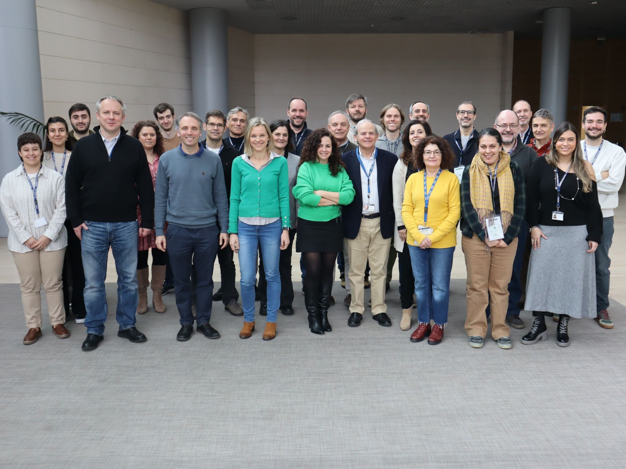
New project (DIGIMATER-CM) in BCD group was starting
Dr. Monica Echeverry-Rendon participated in the kick-off meeting for the DIGIMATER-CM project hosted at IMDEA Materials Institute.

Dr. Monica Echeverry-Rendon participated in the kick-off meeting for the DIGIMATER-CM project hosted at IMDEA Materials Institute.
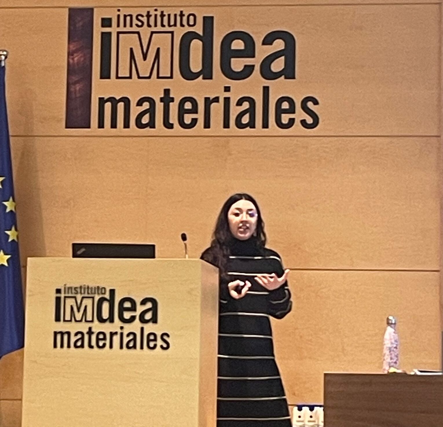
Ángela García de la Camacha Díaz presented her First Year Assessment entitled “Design and fabrication of multimaterial bioresorbable scaffolds by 3D printing for osteochondral tissue regeneration”. She received a positive evaluation
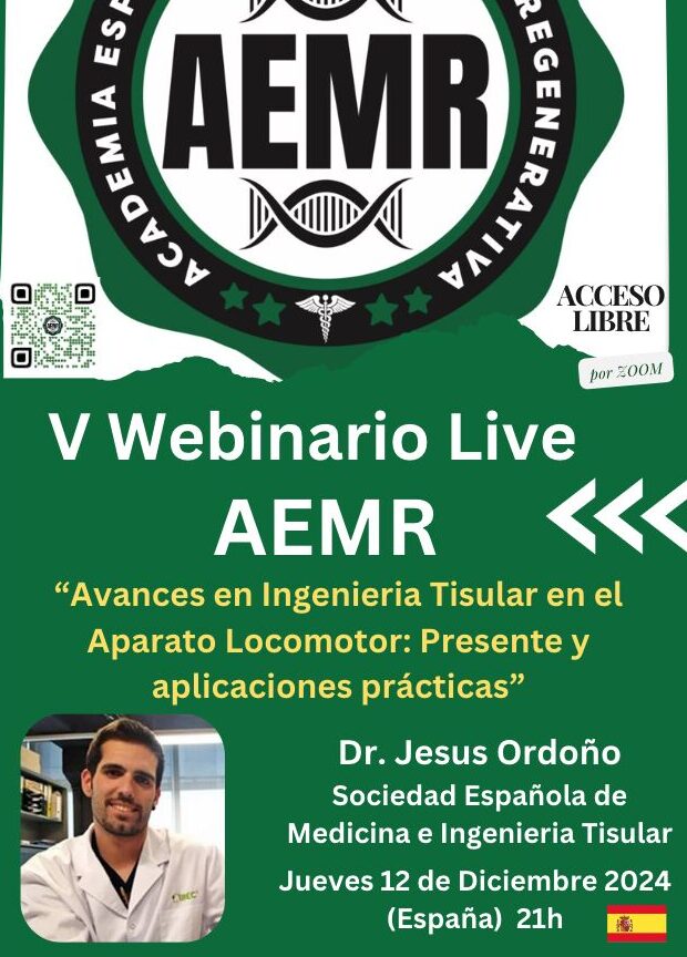
Dr. Jesús Ordoño was invited to give a talk in the V Webinar of AEMR, entitled “Advances in tissue engineering in the musculoskeletal system: present and practical applications.”
Dr. Monica Echeverry-Rendon participated in the kick-off meeting for the DIGIMATER-CM project hosted at IMDEA Materials Institute.
Ángela García de la Camacha Díaz presented her First Year Assessment entitled “Design and fabrication of multimaterial bioresorbable scaffolds by 3D printing for osteochondral tissue regeneration”. She received a positive evaluation
Dr. Jesús Ordoño was invited to give a talk in the V Webinar of AEMR, entitled “Advances in tissue engineering in the musculoskeletal system: present and practical applications.”