Dr. Mónica Echeverry-Rendón Celebrated in CSIC Ranking
We are proud to highlight Dr. Mónica Echeverry Rendón, leader of the BCD Research Group, who has been recognised among Spain’s leading female researchers. The distinction comes from a recently


We are proud to highlight Dr. Mónica Echeverry Rendón, leader of the BCD Research Group, who has been recognised among Spain’s leading female researchers. The distinction comes from a recently
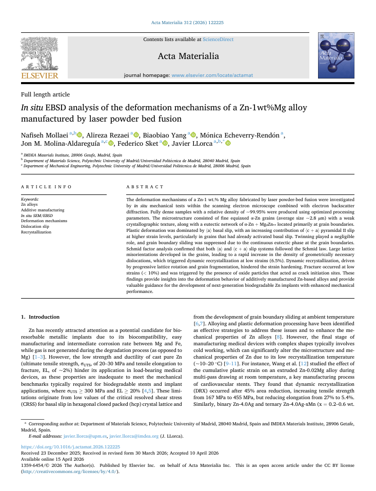
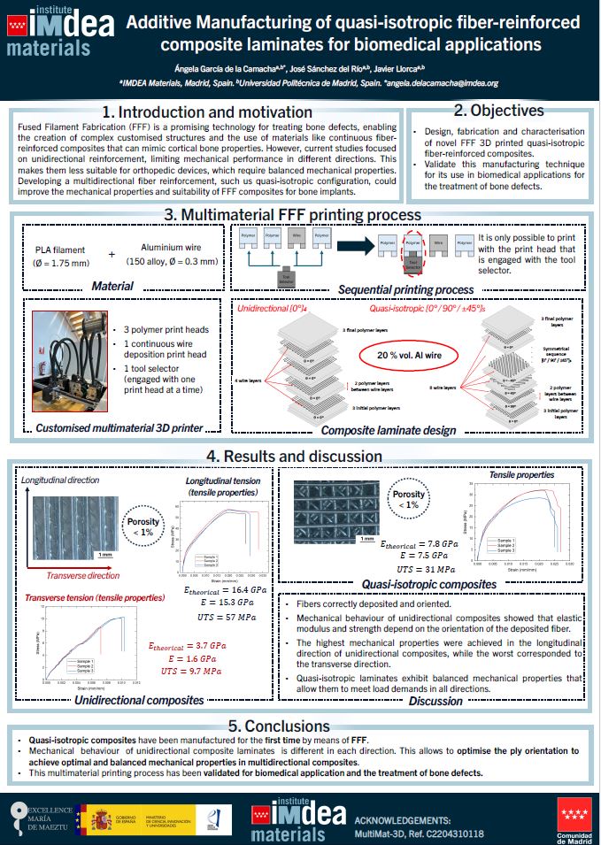
The BCD Group is proud to share our latest publication in Acta Materialia! Congratulations to our colleagues Nafiseh Mollaei, Alireza Rezaei, Biaobiao Yang, Mónica Echeverry-Rendón, Jon M. Molina-Aldreguía, Federico Sket and
The BCD Group is proud to share our latest publication in Antibiotics! Congratulations to our colleagues Bruno F. Gomes-Ribeiro and Mónica Echeverry-Rendón for this excellent achievement! Title: Inhibition of Biofilm
We are proud to highlight Dr. Mónica Echeverry Rendón, leader of the BCD Research Group, who has been recognised among Spain’s leading female researchers. The distinction comes from a recently
The BCD Group is proud to share our latest publication in Acta Materialia! Congratulations to our colleagues Nafiseh Mollaei, Alireza Rezaei, Biaobiao Yang, Mónica Echeverry-Rendón, Jon M. Molina-Aldreguía, Federico Sket and
The BCD Group is proud to share our latest publication in Antibiotics! Congratulations to our colleagues Bruno F. Gomes-Ribeiro and Mónica Echeverry-Rendón for this excellent achievement! Title: Inhibition of Biofilm