3D tissue models can be used in vitro (in the lab) for the study of diseases and the testing of novel drugs, including their incorporation into microfluidic devices to form the so-called “organ-on-chip” systems. 3D tissue models can also be used in vivo (i.e. implanted in the body) to directly promote tissue regeneration and treat damaged or diseased tissues.
As an example of the former, IMDEA Materials is developing more advanced airway-on-chip models that better simulate the airway epithelium and stroma.
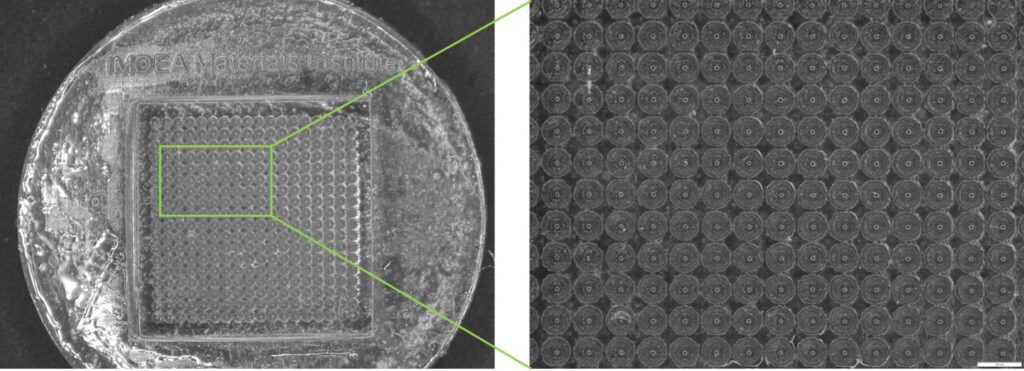
As an example of the latter, our Institute is also studying devices for the formation of organoids (spheroid shaped structures that mimic the natural organization of cells within tissues) using high-resolution 3D printing technologies such as stereolithography (see Figure 1).
We apply our comprehensive know-how in additive manufacturing technologies and 4D characterization in both cases.