
- Within the broad field of advanced therapies, which includes cell therapy and gene therapy, tissue engineering has emerged as one of the most promising tools for transforming the medicine of the future.
Published by: Dr. Pedro J. Díaz Payno. PhD in Biomedical Engineering.
The original version of this article can be found in Spanish in Fundación Muy Interesante.
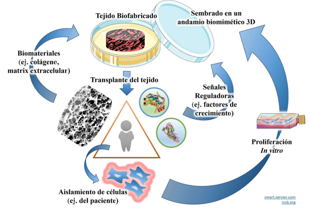
The goal of tissue engineering is to repair, replace or regenerate damaged tissues or organs, whether as a result of disease or injury. Unlike traditional treatments, tissue engineering does not simply aim to relieve symptoms but to restore the original function of damaged tissue. Strategies for tissue regeneration can be broadly divided into two approaches:
- Manufacturing a tissue or organ in the laboratory so that it is ready for transplantation or can serve as a disease model.
- Designing an initial scaffold that, once implanted, matures within the body.
In both strategies, researchers rely on a triad of key components: living cells, which act as the functional units of the tissue; physicochemical regulatory signals, which guide cellular behaviour; and biomaterials, which provide structural support for the cells [1].
Tissue engineering is therefore a multidisciplinary field that combines biology, engineering and materials science. It forms part of the broader concept of regenerative medicine, which seeks to harness the body’s natural capacity for healing through specialised fields such as biofabrication. Thanks to these advances, it is now possible to produce tissues such as skin, cartilage and blood vessels in the laboratory, while moving closer to more complex applications such as organ regeneration.
Cells: The Workforce That Rebuilds the Body
No tissue can regenerate without cells. They are responsible for building, repairing and maintaining the human body [4]. Among the most widely used cell types are mesenchymal stem cells (MSCs), which are capable of producing a variety of tissues, including cartilage, bone and adipose tissue, under laboratory conditions. Their widespread use has led to implantation studies in both animals and humans, although the success of these cell-based therapies has remained limited. As a result, the true nature of MSCs continues to be debated. Many researchers question whether they genuinely meet the classical definition of stem cells. Indeed, Arnold Caplan, who originally coined the term, has proposed redefining them as “Medicinal Signalling Cells” [5], placing greater emphasis on their paracrine function. This reflects how our understanding of these cells has evolved beyond their original definition.
Another highly relevant cell type is endothelial progenitor cells. Endothelial cells contribute to postnatal vasculogenesis, the formation of new blood vessels in adult tissues. These blood vessels are particularly important because they allow newly engineered tissues to integrate successfully with the body and prevent them from becoming deprived of nutrients after implantation.
Other stem cell types include neural stem cells, which have potential applications in developing disease models for neurodegenerative disorders such as Alzheimer’s disease and amyotrophic lateral sclerosis (ALS), as well as corneal stem cells for regenerating the ocular surface and corneal stroma. Adult cells also offer significant potential in regenerative medicine. Fibroblasts, for example, can be used to engineer skin, while chondrocytes (cartilage cells) are valuable for repairing joints such as the knee.
Finally, one of the most remarkable advances of recent years has been the development of induced pluripotent stem cells (iPSCs). Scientists have learned how to “rewind” adult cells to an earlier developmental state, restoring their ability to generate almost any tissue type. In tissue engineering, this raises the possibility that every patient could become their own donor, opening the door to truly personalised therapies.
The biological traffic signals that guide regeneration
Cells do not act alone. They require physicochemical signals that instruct them when to grow, migrate, divide or differentiate. In many ways, these signals function like biological traffic lights, coordinating cellular behaviour.
On the one hand, there are physical signals, including mechanical stimuli generated by bioreactors or by the intrinsic properties of the biomaterial itself. Bioreactors can reproduce forces naturally experienced within the body, such as the tension and compression exerted on our joints during walking. Likewise, properties of the supporting biomaterial, including stiffness, topography and elasticity, can significantly influence how cells behave.
Chemical and biochemical signals include molecules capable of activating specific metabolic pathways. Among these are growth factors found within the extracellular matrix of many tissues [7], which play essential roles in processes such as cell proliferation and differentiation. Other regulatory molecules, such as insulin, together with chemical gradients that guide cellular behaviour through space, are equally important.
Biomaterials: The LEGO Bricks of Regenerative Medicine
If cells are the “workers” and signals are the “instructions”, biomaterials provide the platform upon which new tissues are built. In many ways, they function like biological LEGO bricks, allowing cells to organise themselves into complex structures. Essentially, they act as scaffolds that guide tissue regeneration.
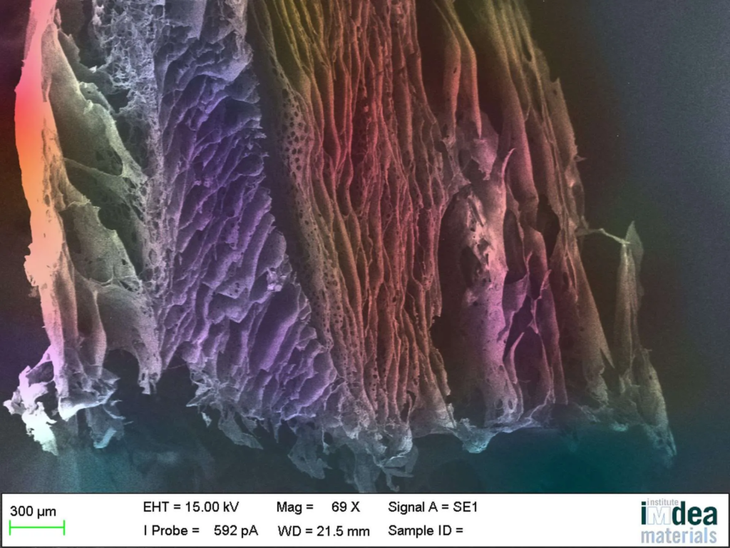
Not just any material will do. A biomaterial must be biocompatible, meaning it is non-toxic and does not harm the body. Ideally, it should also be biodegradable, gradually disappearing as the tissue regenerates. At the same time, it must enable cells to attach, migrate and respond to biological signals while reproducing, as closely as possible, the body’s natural environment.
There are many different classes of biomaterials. Some are naturally derived, such as collagen, fibrin and hyaluronic acid, which closely resemble the extracellular matrix of human tissues [8]. Others are synthetic, including polymers such as PLA, PCL and PEG, ceramics and even metals, which are designed to provide specific properties such as mechanical strength or controlled degradation. The most advanced approaches seek to combine the advantages of both natural and synthetic materials, creating scaffolds that are both mechanically robust and biologically active.
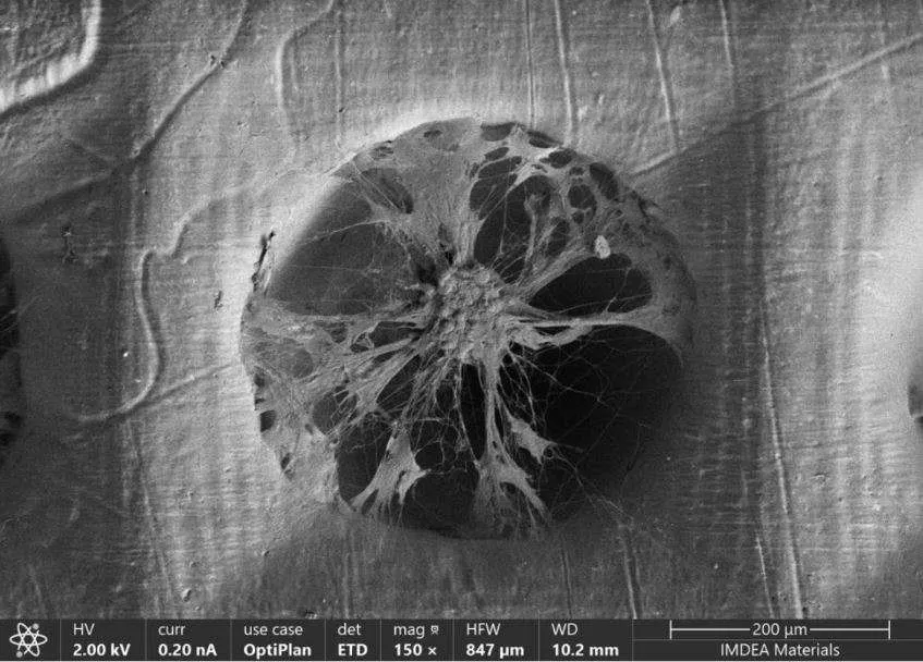
The methods used to manufacture these biomaterials are also becoming increasingly sophisticated. Advanced manufacturing techniques such as 3D printing make it possible to design structures with unprecedented precision, controlling characteristics such as porosity, geometry and the distribution of multiple materials within a scaffold. This enables the creation of structures that more closely resemble native tissues. In the case of 3D bioprinting, biomaterials and living cells can be deposited layer by layer, bringing increasingly complex functional tissues within reach.
This evolution in materials and manufacturing technologies illustrates how medicine itself has changed. Early biomaterials were essentially inert, serving only to replace a missing function without interacting with the body. Today’s biomaterials, by contrast, are dynamic structures designed to communicate with cells, release biological signals and even respond to external stimuli. These so-called smart materials, or 4D materials [9], actively promote tissue regeneration throughout the healing process.
Far More Than Tissue Repair: Mini-Organs, Cancer and the Medicines of Tomorrow
Tissue engineering is not only transforming how we repair the human body. It is also changing the way diseases are studied and new medicines are developed. One of its most promising applications is the creation of more realistic laboratory models of disease, which are essential for understanding the progression of complex conditions such as cancer and for improving the success rate of new drugs and treatment regimens before they reach patients.
This also offers a significant ethical and scientific advantage by helping to reduce the use of animals in research, one of today’s major priorities. One of the greatest challenges in biomedical science is that treatments which perform well in animal models often fail to achieve the same results in humans. This highlights the need for more advanced and reliable experimental systems capable of bridging the gap between laboratory research and clinical reality. A striking example is paracetamol: while safe for humans, it is toxic to cats. Had this medicine been evaluated primarily in feline preclinical models, it would probably never have been approved. It serves as a clear reminder that different species can respond very differently to the same treatment.
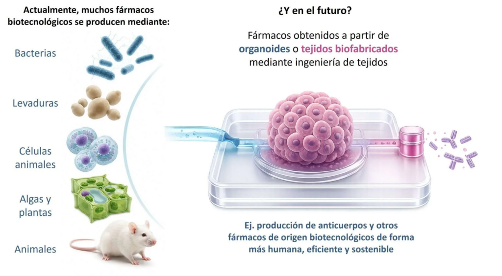
Beyond tissue regeneration, tissue engineering could also transform the production of complex biopharmaceuticals. Many medicines are currently manufactured using bacteria, yeast or mammalian cell lines, such as the well-known CHO cells. However, certain particularly complex molecules, including some forms of heparin, still depend on animal tissues, primarily of porcine origin. Even low-molecular-weight heparins, which offer more precise control of blood coagulation, continue to rely on animal-derived sources. In this context, the development of mini-organs or organ-on-a-chip systems capable of producing these compounds under controlled conditions represents far more than an incremental improvement. It marks a genuine paradigm shift, reducing dependence on animal sources while enabling drug production in systems that more closely replicate human physiology.
From bioprinters to the marketplace: Spain’s race to keep pace
The race to manufacture human tissues is no longer confined to universities and hospitals. Increasingly, companies are seeking to transform scientific advances into commercial products and technologies.
In Spain, companies such as REGEMAT3D have helped place the country on the international biofabrication map through the development of their own internationally recognised bioprinters. Alongside them, new ventures such as Health Biolux are emerging, specialising in DLP (Digital Light Processing)-based bioprinters that use light to fabricate structures with extremely high resolution and architectural complexity. These technologies make it possible to reproduce more realistic tissues by organising biomaterials and living cells with exceptional precision.
Other notable companies include Technical Proteins Nanobiotechnology and Silk Biomed, which specialises in protein-based biomaterials for tissue regeneration. Together, they demonstrate how laboratory discoveries are beginning to translate into tangible biomedical solutions.
The ecosystem also includes companies from unexpected sectors. One striking example is Viscofan Bioengineering, a division of the well-known manufacturer of sausage casings, which has successfully transferred its expertise in collagen processing to the development of biomaterials for biomedical applications. It illustrates how technologies developed far from medicine can find entirely new opportunities in regenerative healthcare.
Although Spain’s ecosystem is still some distance from matching the competitiveness of leading countries such as the United States, Germany and the Netherlands, its potential is increasingly evident. A strong scientific foundation, the steady growth of innovative companies and initiatives such as the Spanish Society for Regenerative Medicine and Tissue Engineering (SEMIT), which aims to connect researchers, clinicians and industry, all demonstrate that the field is reaching maturity [10].
Spain may not yet be leading this global race, but it is steadily building the foundations needed to ensure it does not fall behind.
References
- Langer R, Vacanti JP. Tissue engineering. Science. 1993 May 14;260(5110):920-6. doi: 10.1126/science.8493529
- https://smart.servier.com
- https://www.rcsb.org/
- Vunjak-Novakovic G, Freshney RI, editors. Culture of Cells for Tissue Engineering. Hoboken (NJ): John Wiley & Sons, Inc.; 2005. doi:10.1002/0471741817
- Caplan AI. Mesenchymal Stem Cells: Time to Change the Name! Stem Cells Transl Med. 2017 Jun;6(6):1445-1451. doi: 10.1002/sctm.17-0051
- Navarrete-Segado P, et al. High-throughput 3D spheroid production with photocurable polyurethane microwell arrays via LCD 3D printing. ChemRxiv. 2019. doi:10.26434/chemrxiv.10001840.v1
- Díaz-Payno PJ, et al. The identification of articular cartilage and growth plate extracellular matrix-specific proteins supportive of either osteogenesis or stable chondrogenesis of stem cells. Biochem Biophys Res Commun. 2020;530(3):503–509. doi:10.1016/j.bbrc.2020.05.074
- Browe DC, Díaz-Payno PJ, et al. Bilayered extracellular matrix derived scaffolds with anisotropic pore architecture guide tissue organization during osteochondral defect repair. Acta Biomater. 2022;143:266–281. doi:10.1016/j.actbio.2022.03.009
- Yarali E, et al. 4D printing for biomedical applications. Adv Mater. 2024:e2402301. doi:10.1002/adma.202402301.
- https://semit.es/
The original version of this article can be found in Spanish in Fundación Muy Interesante.