
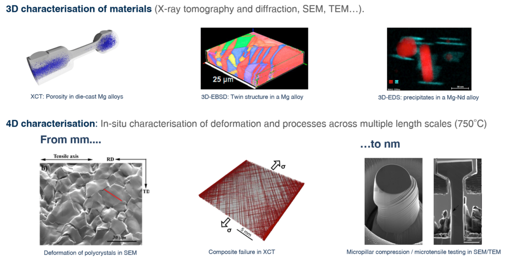
Progress in the development of new materials and processing methods can only come from a thorough understanding of the microstructure of the material in focus, its evolution during either processing or service operation, and its influence on the relevant properties for the purpose it was designed. Since the microstructural features that determine material behaviour usually span several length scales for instance, from macroscopic defect distribution to nanometre scale precipitates in the case of metallic alloys, this understanding can only come from advanced 4D characterisation techniques, capable of determining the evolution of the 3D microstructure over time at different length scales; hence the term 4D. This is precisely the

objective of this programme: to understand microstructure/defect distribution and evolution in advanced materials during processing and service using advanced characterisation techniques.
Necessary cookies are absolutely essential for the website to function properly. This category only includes cookies that ensures basic functionalities and security features of the website. These cookies do not store any personal information.
| Cookie | Type | Duration | Description |
|---|---|---|---|
| cookielawinfo-checkbox-advertisement | persistent | 1 year | This cookie is set by GDPR Cookie Consent plugin. The purpose of this cookie is to check whether or not the user has given their consent to the usage of cookies under the category 'Advertisement'. |
| cookielawinfo-checkbox-analytics | persistent | 1 year | This cookie is set by GDPR Cookie Consent plugin. The purpose of this cookie is to check whether or not the user has given the consent to the usage of cookies under the category 'Analytics'. |
| cookielawinfo-checkbox-necessary | persistent | 11 months | This cookie is set by GDPR Cookie Consent plugin. The cookies is used to store the user consent for the cookies in the category "Necessary". |
| cookielawinfo-checkbox-non-necessary | persistent | 11 months | This cookie is set by GDPR Cookie Consent plugin. The cookies is used to store the user consent for the cookies in the category "Non Necessary". |
| cookielawinfo-checkbox-performance | persistent | 1 year | This cookie is set by GDPR Cookie Consent plugin. The purpose of this cookie is to check whether or not the user has given the consent to the usage of cookies under the category 'Performance'. |
| PHPSESSID | persistent | 1 year | This cookie is native to PHP applications. The cookie is used to store and identify a users' unique session ID for the purpose of managing user session on the website. The cookie is a session cookies and is deleted when all the browser windows are closed. |
| viewed_cookie_policy | persistent | 11 months | The cookie is set by the GDPR Cookie Consent plugin and is used to store whether or not user has consented to the use of cookies. It does not store any personal data. |
Analytical cookies are used to understand how visitors interact with the website. These cookies help provide information on metrics the number of visitors, bounce rate, traffic source, etc.
| Cookie | Type | Duration | Description |
|---|---|---|---|
| _ga | persistent | 2 years | This cookie is installed by Google Analytics. The cookie is used to calculate visitor, session, camapign data and keep track of site usage for the site's analytics report. The cookies store information anonymously and assigns a randoly generated number to identify unique visitors. |
| _gid | persistent | 1 day | This cookie is installed by Google Analytics. The cookie is used to store information of how visitors use a website and helps in creating an analytics report of how the wbsite is doing. The data collected including the number visitors, the source where they have come from, and the pages viisted in an anonymous form. |
| GPS | persistent | 30 minutes | This cookie is set by Youtube and registers a unique ID for tracking users based on their geographical location |
Performance cookies are used to understand and analyze the key performance indexes of the website which helps in delivering a better user experience for the visitors.
| Cookie | Type | Duration | Description |
|---|---|---|---|
| _gat | persistent | 1 minute | This cookies is installed by Google Universal Analytics to throttle the request rate to limit the colllection of data on high traffic sites. |
| YSC | 1 | 1 year | This cookies is set by Youtube and is used to track the views of embedded videos. |
Advertisement cookies are used to provide visitors with relevant ads and marketing campaigns. These cookies track visitors across websites and collect information to provide customized ads.
| Cookie | Type | Duration | Description |
|---|---|---|---|
| IDE | 1 | 2 years | Used by Google DoubleClick and stores information about how the user uses the website and any other advertisement before visiting the website. This is used to present users with ads that are relevant to them according to the user profile. |
| VISITOR_INFO1_LIVE | 1 | 5 months | This cookie is set by Youtube. Used to track the information of the embedded YouTube videos on a website. |